The Retinal Pigment Epithelium and Its Role in AMD
July 8, 2026

What the RPE Does in Your Eye
The RPE sits between the light-sensing cells of the retina (called photoreceptors) and the blood vessels beneath them, known as the choroid. This location makes the RPE one of the hardest-working tissue layers in the entire body, responsible for keeping the photoreceptors alive and functioning every moment of your life.
The RPE is a single layer of hexagonal, pigment-rich cells. Its upper surface connects directly to the photoreceptors through tiny projections called microvilli, and its lower surface rests on a thin membrane called Bruch's membrane, which separates it from the choroidal blood supply below.
This position means every nutrient your photoreceptors need must pass through the RPE first. Every waste product the photoreceptors generate must be cleared by the RPE in return.
RPE cells carry out several functions that are critical to sustained, clear vision. Without them, the photoreceptors would deteriorate quickly.
- Daily cleanup of worn-out photoreceptor material through a process called phagocytosis
- Recycling of 11-cis-retinal, the light-sensitive molecule photoreceptors need to detect light (this is called the visual cycle)
- Transporting nutrients such as glucose and fatty acids from the blood supply to the photoreceptors
- Absorbing excess light through melanin pigment to protect the retina from light-related oxidative damage
- Secreting growth factors that maintain the health of the choroidal blood vessels beneath them
- Maintaining the outer blood-retinal barrier, which controls what enters the retinal space from the bloodstream
Because RPE cells do not divide or regenerate in adults, the same cells you had as a young adult are the ones your eyes rely on for the rest of your life.
Over decades of daily phagocytosis, the internal machinery of RPE cells, particularly the lysosomes that digest waste, gradually becomes less efficient. Incompletely digested material builds up inside the cells as a toxic pigment called lipofuscin.
This accumulation is not just a sign of aging. Lipofuscin actively damages RPE cells from within, setting the stage for AMD.

How RPE Breakdown Drives Dry AMD
Dry AMD develops through a sequence of RPE changes that unfold over years or even decades. Each step places greater stress on the RPE and the photoreceptors that depend on it. Understanding this sequence helps explain why early detection matters so much.
As lipofuscin builds up inside RPE cells, a component of it called A2E generates toxic molecules called reactive oxygen species when exposed to light. These molecules damage cell membranes and DNA from within, further impairing the RPE's ability to do its job.
Your doctor can detect lipofuscin buildup using a painless imaging technique called fundus autofluorescence. Areas with high autofluorescence on these images indicate RPE cells that are stressed and heavily loaded with lipofuscin. These areas often show where future damage is most likely to develop.
When RPE cells cannot export waste efficiently through Bruch's membrane, that waste accumulates between the RPE and the membrane below, forming deposits called drusen. Small drusen can appear as a normal part of aging, but larger or more numerous drusen are a warning sign that the RPE is struggling in that area.
As drusen grow, they physically lift the RPE away from its choroidal blood supply, cutting off oxygen and nutrients. This creates a damaging cycle: RPE stress leads to drusen, and drusen worsen RPE stress, increasing the likelihood that cells will die.
When enough RPE cells die in a concentrated area, the photoreceptors directly above them also degenerate because they can no longer survive without RPE support. The resulting area of cell death is called geographic atrophy (GA), which represents the advanced form of dry AMD.
The progression typically follows a predictable pattern: drusen form first, RPE cells show signs of stress and pigment changes, and then RPE loss and atrophy follow. Tracking this pattern at regular eye appointments allows your doctor to predict where atrophy may spread and guide treatment decisions accordingly.

RPE Dysfunction and Wet AMD
While dry AMD results from the slow death of RPE cells, wet AMD develops when RPE dysfunction triggers the growth of abnormal blood vessels that leak fluid into the retina. Both forms share the same underlying RPE breakdown, but wet AMD can cause rapid vision changes and requires timely treatment.
Healthy RPE cells release controlled amounts of a protein called vascular endothelial growth factor (VEGF), which helps maintain the normal choroidal blood vessels beneath the retina. When RPE cells are damaged or stressed, they can release excess VEGF.
This excess VEGF stimulates the growth of abnormal new blood vessels, a process called choroidal neovascularization. These vessels are fragile and tend to leak fluid or blood into the retinal layers, causing rapid and sometimes severe vision loss. Anti-VEGF injections, the current standard treatment for wet AMD, work by blocking these excess growth signals.
We use several imaging tools to assess the health of your RPE layer at each visit. Each test reveals different aspects of RPE function and structure.
- Optical coherence tomography (OCT) shows the thickness, elevation, and integrity of the RPE layer in cross-section
- Fundus autofluorescence reveals areas of lipofuscin accumulation (appearing bright) and areas of RPE cell loss (appearing dark)
- OCT angiography can detect abnormal blood vessel growth beneath the retina before it causes noticeable symptoms
Together, these tests give us a detailed map of your RPE across the macula, the central region of the retina responsible for sharp, detailed vision. This map guides every treatment and monitoring decision we make for you.
The stage of RPE health revealed by imaging directly determines which treatments are appropriate. When the RPE is stressed but still intact, measures such as AREDS2 nutritional supplements and protective lifestyle changes may help slow further damage.
When geographic atrophy has already formed, newer complement-targeting therapies may help slow its expansion. When excess VEGF from dysfunctional RPE has triggered wet AMD, anti-VEGF injections reduce fluid accumulation and protect the remaining retina. Tailoring treatment to the specific state of your RPE is a core part of the care we provide.

Frequently Asked Questions
These answers address common questions about RPE health and AMD that go beyond what is covered in the sections above.
RPE cells have a limited ability to clear accumulated waste and recover from mild, early stress. However, once an RPE cell has died, it is not replaced by the body. The permanent nature of RPE cell loss is exactly why monitoring and early intervention matter so much. Preserving the RPE cells you still have is the central goal of current AMD care.
Stem cell-derived RPE replacement is currently in clinical trials and is not yet an approved standard treatment. Researchers are developing lab-grown RPE cell patches that could potentially be implanted under the retina to replace lost cells. These trials are still in early phases and long-term outcomes are being evaluated, so it is important to discuss any emerging options directly with your doctor rather than seeking unregulated procedures.
UV-blocking sunglasses reduce the amount of light-related oxidative stress reaching the retina, which may lower the cumulative burden on RPE cells over time. While sunglasses alone cannot prevent AMD, reducing photo-oxidative exposure is a reasonable and low-risk protective habit, especially for people who already have drusen or documented RPE changes. Polarized lenses with full UV protection are the best choice for outdoor use.
Nutrients such as lutein and zeaxanthin, found in leafy greens and colorful vegetables, accumulate in the macula and act as natural light filters, reducing the oxidative load on RPE cells. Omega-3 fatty acids from fish may also support retinal health. AREDS2 supplements are specifically formulated to provide antioxidants shown to slow AMD progression in people with intermediate or advanced disease. A healthy diet supports RPE health but does not replace regular monitoring and medical care.
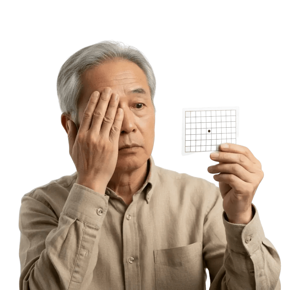
Sudden changes such as new distortion, a dark or blurry spot in your central vision, or straight lines appearing wavy can indicate wet AMD developing or progressing. These changes require prompt evaluation, often within one to two days, because early treatment of wet AMD protects more vision. Do not wait for your next scheduled appointment if you notice any sudden shift in vision quality.
Monitoring frequency depends on how advanced your AMD is and how stable your RPE appears on imaging. People with early AMD may need annual visits, while those with intermediate AMD or geographic atrophy often benefit from visits every three to six months. If you have wet AMD being treated with injections, your doctor will set a schedule based on how your retina responds. Consistent follow-up is the most reliable way to catch changes before they cause irreversible vision loss.
We Are Here to Help You Protect Your Vision
The RPE is a remarkable tissue that works silently and continuously to keep your photoreceptors alive and your vision clear. Our team is experienced in detecting and managing every stage of RPE-related disease, from early drusen to geographic atrophy and wet AMD. We use advanced imaging at every visit to stay ahead of changes and tailor your care to exactly what your retina needs. If you have concerns about AMD or your retinal health, we encourage you to schedule a comprehensive evaluation with us.